Photography has revolutionized dentistry, just as it has changed many other industries. Today, dentists are improving patient care by capturing highly detailed images of the inside of the mouth.
Understandably, this leads to more accurate diagnosis, treatment planning, and monitoring of dental conditions.
Dr. Elizabeth C. Robinson on Rock Hill WRHI Radio: Dental Photography (2 minutes)

Best of all, patients can see exactly what their teeth and gums look like and where dentists see problem areas.
In this blog post, I will discuss how photography and intraoral photos are helpful for our dentists, hygienists, and patients.
Dental Photography
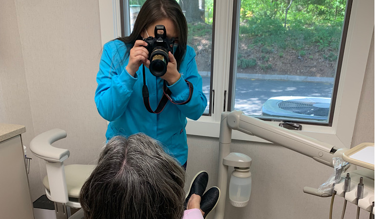
Photography is an essential tool at Cranford Dental. Dr. Cranford began using intraoral dental photography in the early 1990s– long before it became industry standard. In those days, he had a larger camera and screen.
Today the intraoral camera is very tiny. We project the images onto our TV screen for the patient to see.
Cameras used in Dentistry
Cameras used in dental offices take very precise photos. In fact, the photos give a clearer picture of the details of the teeth than looking into the mouth gives the patient and dentist.
At Cranford Dental, we use both Intraoral and Digital SLR Cameras to improve the patient experience.
Intraoral Cameras
Intraoral cameras are small and designed to fit into the patient’s mouth.
These cameras link into our computer system so that the patient in the chair can immediately look at problem areas such as broken teeth, cracks, and decay.
We have small intraoral cameras in each room. Invaluable to our dentists, they take photos of individual teeth or areas of the mouth.

Digital SLR Cameras
We also use a digital SLR camera to take photos which show the whole mouth or smile all at once. Sometimes we use specialized lenses and flashes to show even more detail.
Our dentists and staff have specific training in using cameras to get good close up photos of the outside and inside of the mouth.
Accurate Diagnosis
One of the most significant benefits of intraoral photography is the ability to capture highly detailed images of the mouth. These images can be magnified and zoomed in to provide a clear view of the teeth and gums, allowing our dentists to detect even the smallest changes or abnormalities.
This level of detail is impossible to see with the naked eye.
Intraoral photos help dentists diagnose conditions such as cavities, cracks in the teeth, and oral cancer at an early stage when they are more easily treated.
This intraoral photo shows the dentist and the patient an old filling and fractures in the tooth
Treatment Planning
Intraoral photos are helpful in treatment planning. Once a dental condition is diagnosed, the dentist can use the images to develop a personalized treatment plan. For example, if a patient has a cavity, the dentist can use the images to determine the size and location of the cavity, and decide on the best course of action, such as a filling or crown.
In cosmetic cases, photos show the relationship between the teeth and the rest of the face. The dentist studies photos of the patient to make the decision about the proper placement of veneers and crowns to achieve the most esthetic and functional result.
Photos allow us to spend additional time planning after the patient leaves the office.
When the patient saw her “before” picture, she understood that the proportions of her old veneers were wrong. This helped her understand why she would need to have 4 veneers to make her smile ideal.
We also use photos to check for changes over time. For example, an area of recession in the gums may be less concerning if we know it has been there for a long time.
It is much more alarming if something develops and changes very rapidly. Photos help determine how aggressively we need to treat conditions such as recession and erosion.
I can’t imagine practicing dentistry without the use of photos. They improve the experience for both the dentist and the patient. Diagnosis and planning is much clearer when we have the proper photographs.
Dr. Elizabeth C. Robinson
Patient Education
We use photos to educate our patients about their dental health. Seeing detailed images of teeth and gums helps patients better understand their condition and the plan to fix it.
In most instances you don’t have to just take your dentist’s word for it that something is wrong– you can see it for yourself! Your dentist or hygienist will use photos, xrays, and models to educate you.
You shouldn’t move forward with any dental treatment that you don’t understand.
Your dentist also uses intraoral photos to demonstrate the effectiveness of treatment, showing patients the progress they have made over time. Showing patients “before and after” photos helps them understand what we are able to achieve with proper dental treatment.
This patient had never had a cavity before, so he was skeptical about accepting treatment. These photos of the cavity before and during treatment helped him understand why he needed a filling.
Improved Communication
Intraoral photos can also improve communication between dental professionals. By sharing images with other specialists or referring dentists, dental professionals can collaborate more effectively. This ensures that the patient receives the best possible care.
Intraoral photos and DSLR photos help communicate with dental laboratories. This allows the lab to make restorations that match the patient’s natural teeth.
Photos also help communicate with insurance companies to help our patients get proper coverage. Kathy and Lisa submit photos of your teeth so that the insurance company will understand why we completed certain treatments.
Insurance companies want to see evidence that a tooth needs a crown. This photo shows a large metal filling with decay around it. Without a crown, this tooth is likely to fracture.
What to Expect at Cranford Dental
At your first visit with Cranford Dental we take a full series of x-rays and photos. Our dentists will use these images to help diagnose any problems and to make a plan for your dental health.
We will show you photos of the teeth that have problems and explain to you what we see.
If you do not currently have any problems with your teeth, the photos are used to look for changes over time. For example, if you come in with a broken filling, we will be able to look back at pictures taken years earlier to see what the condition of the filling was at that time.
If you are interested in cosmetic treatment or have more complicated dental needs, we may suggest a separate appointment to take additional photos.
We want you to totally understand what your teeth need and why they need it.
We trust that Cranford Dental’s use of dental photography will help you have a better dental experience.
Contact our office in Rock Hill, SC or call 803-324 -7670 if you would like to get a good look at your teeth or the inside of your mouth. We enjoy welcoming new patients to Cranford Dental.
Leave a Reply